Bone augmentation and sinus floor elevation
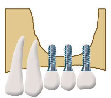
The maxillary sinus lies directly above the alveolar ridge of the upper jaw. After a tooth is removed, or when the posterior upper teeth are lost, the bone recedes. As a result the bone of the alveolar ridge loses height. Often only a thin layer of bone remains in the upper jaw, which is no longer sufficient to hold an endosseous implant. This bone deficit can be compensated for by what is known as sinus floor elevation.
Raising the floor of the maxillary sinus is a special form of bone augmentation. Where there is too little bone in the upper jaw, the sinus can be raised by placing bone or bone substitute material into the maxillary sinus. Where bone is scarce in the posterior region of the upper jaw, a sinus lift is often the only way to create sufficient bone for implant treatment and fixed teeth. When a sinus floor elevation is planned, 3D diagnostics provides detailed information before the procedure about the exact anatomical situation and the extent to which bone needs to be built up.
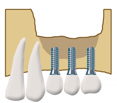
A distinction is made between the internal and the external sinus lift. The external sinus lift is carried out under general anaesthetic: the maxillary sinus is opened surgically through the oral cavity in the region of the cheek so that bone can be introduced under direct vision. In the internal sinus lift, the implant channel - the drilled hole for the implant - is used to place the bone material. This channel is pierced carefully while protecting the sinus membrane, so that the sinus itself remains unopened. The membrane is raised with special instruments called osteotomes, and the augmentation material is then placed through this channel. The additional bone makes it possible to place longer implants straight away and to achieve greater stability with them after healing. It is important that the maxillary sinus is free of infection at the time of the sinus lift operation.