Dental knowledge: digital 3D planning
Layered images produce a three-dimensional picture of tooth and jaw. It allows the position, angle and depth of the implant to be determined in advance.
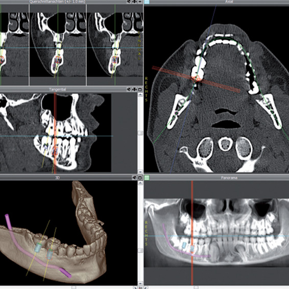
Planning begins with layered images of the jaw. The patient wears a template with markers that reappear on the radiograph. From the imported data a three-dimensional picture of the teeth and jaw emerges.
It allows the ideal seat of the implant to be determined – position, angle and depth – along with precise information on the structure and quality of the bone and on the course of the mandibular nerve and the maxillary sinuses.
What this means for safety
Because the anatomy is fully known before the procedure, the plan can be aligned with the actual conditions. The risk of injuring tooth roots, neighbouring teeth, the maxillary sinus or nerves drops considerably.